What happens in the body
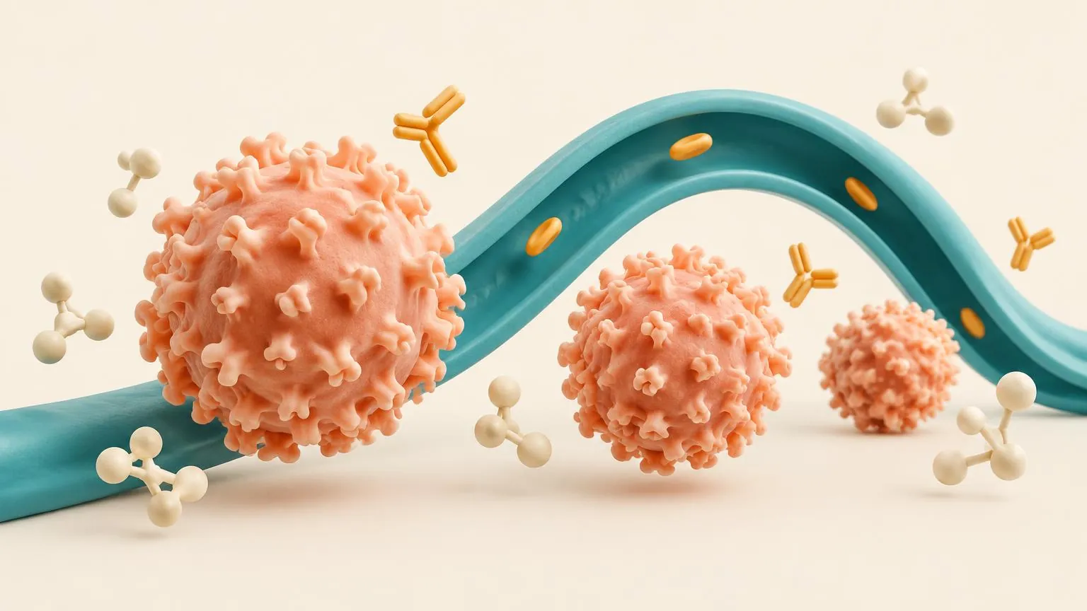
The immune system turns on the nervous system
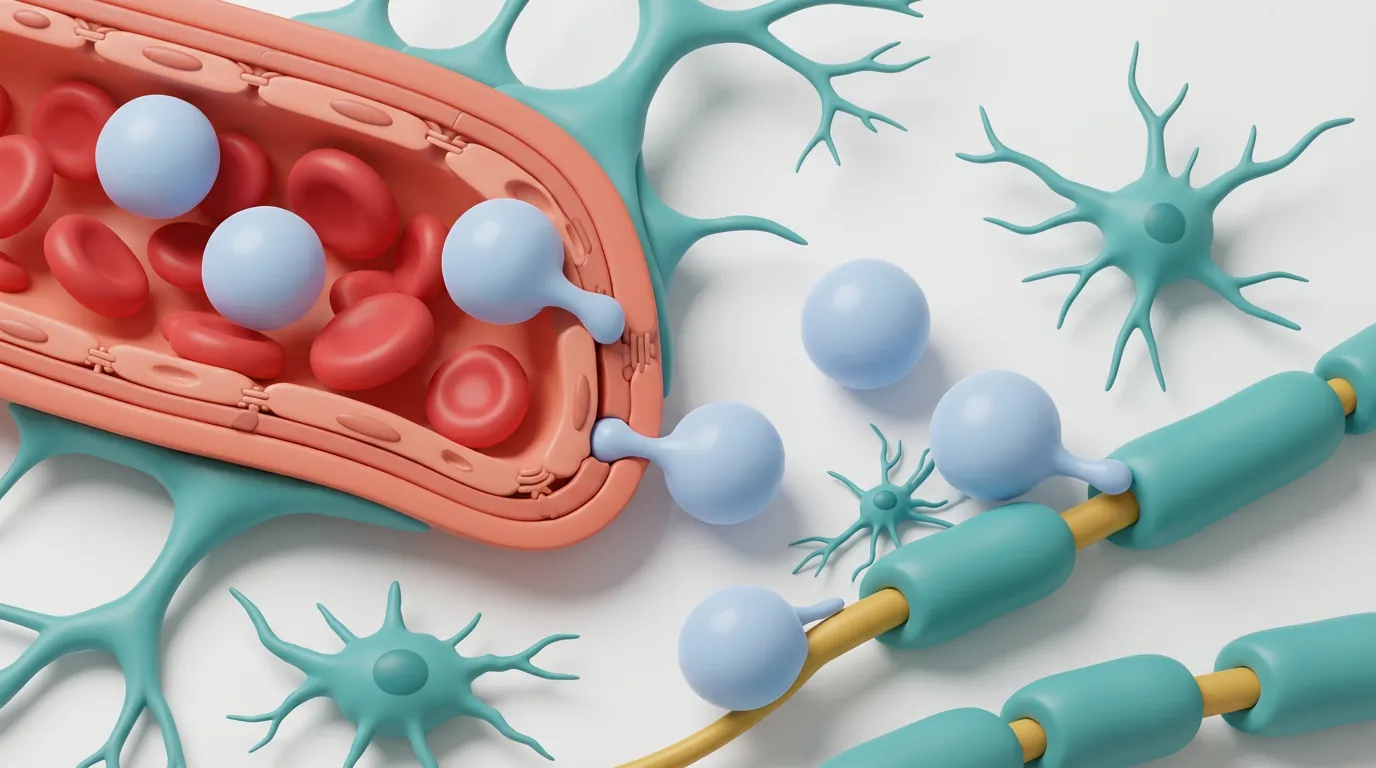
The brain and spinal cord are normally sheltered from most of the immune system by the blood-brain barrier, a tight lining of the small blood vessels that supply them. In MS, immune cells that have been activated elsewhere in the body cross this barrier and set up inflammation inside the nervous system. Their main target is myelin and the cells that make it, called oligodendrocytes.
For many years MS was thought to be driven mainly by T cells, partly because the animal model used to study it is a T-cell disease. In 2008 a trial of rituximab, a medicine that removes B cells, showed a large and rapid fall in new inflammation, and B cells have been at the centre of MS research ever since.
Why the immune system makes this mistake is not fully known. One explanation under study is molecular mimicry: in some people with MS, antibodies against a protein of the Epstein-Barr virus also bind GlialCAM, a protein found in the brain.
An attack, step by step
Step 1 of 5 · Activation
Lesions
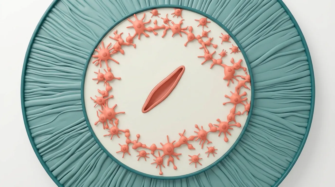
Where inflammation strikes, it leaves a lesion, an area where myelin has been stripped away and the tissue scarred. Lesions are easiest to see on MRI in the white matter, the bundles of nerve fibres deep in the brain and in the spinal cord. They also form in the grey matter of the cortex, often from the earliest stages of the disease, although standard MRI shows these poorly.
Nerve fibres, not only myelin
Losing myelin slows or blocks the signals that a nerve fibre carries. The fibres themselves are also cut in active lesions, and studies of brain tissue have found large numbers of severed fibres in lesions at every stage of the disease. A nerve fibre that is lost is not replaced, and this loss is thought to explain much of the disability that builds up over time.
The brain repairs itself, up to a point
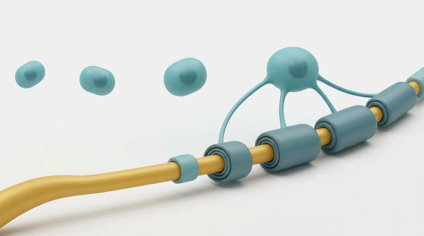
The brain can make new myelin. Precursor cells move into a lesion, mature into oligodendrocytes and wrap the bare fibres again, a process called remyelination. In some people this repair is extensive, while in others most lesions stay bare.
Repair often stalls at a late step. In long-standing lesions, researchers have found immature oligodendrocytes lying next to bare fibres, apparently unable to finish the job. The capacity for repair also declines with age, which experiments in animals link to the ageing of the precursor cells themselves.
Smouldering inflammation
Not all of the inflammation in MS comes from cells arriving from the blood. Some lesions stay active at their edges for years, with microglia, the brain's own immune cells, slowly extending the damage. Clusters of B cells can also form in the membranes that cover the brain, close to areas of damaged cortex.
This inflammation sits behind the blood-brain barrier, where many medicines that act on immune cells in the blood reach poorly. It is one of the leading explanations for why disability can keep worsening after relapses have been brought under control.
Reserve
The brain can compensate for damage by rerouting signals and recruiting other areas. This reserve helps explain why the lesions on a scan and a person's symptoms often do not match, and why disability can appear quite suddenly once the reserve has been used up. Reserve falls with age.
In more depth
Inflammation and nerve damage go together
In a study of 67 brains from people with MS at different stages, injury to nerve fibres went hand in hand with inflammation in every stage, including the progressive stages. This is one of the findings behind the current view that inflammation and neurodegeneration are two sides of one process rather than separate phases. In some older people with long-standing disease, inflammatory infiltrates had fallen to levels similar to those in controls.
Common questions
In the research now
Loading current research…
Key terms
- Myelin
- The fatty sheath wrapped around many nerve fibres in the brain and spinal cord. It lets signals jump quickly along the fibre. MS damages it.
- Axon (nerve fibre)
- The long extension of a nerve cell that carries its signal. Losing axons, rather than myelin alone, is thought to cause most lasting disability.
- Oligodendrocyte
- The cell in the brain and spinal cord that makes myelin. One oligodendrocyte can wrap many axons.
- Remyelination
- The brain's own repair of lost myelin, by new oligodendrocytes. It happens in MS but often falls short.
- Blood-brain barrier
- The tight lining of the small blood vessels of the brain and spinal cord, which keeps most immune cells and large molecules out.
- T cell
- A white blood cell that recognises a specific target and directs or carries out an immune attack.
- B cell
- A white blood cell that can present targets to T cells, release inflammatory signals and mature into antibody-making cells. Some B-cell-targeting medicines are highly effective MS treatments.
- Microglia
- The resident immune cells of the brain and spinal cord. In MS they sustain slow inflammation at the edges of chronic lesions.
- Lesion (plaque)
- An area of damage where myelin has been lost and the tissue scarred.
- Relapse (attack)
- New or worsening symptoms lasting at least 24 hours, without fever or infection to explain them.
- Epstein-Barr virus (EBV)
- A common herpes virus that causes glandular fever. Infection appears to be almost necessary for MS, but most infected people never develop it.
- Molecular mimicry
- When the immune response to a microbe also recognises a similar-looking protein of the body.
References
- NINDS · Multiple sclerosis ↗
Primary research
- Hauser et al. · B-cell depletion with rituximab in relapsing-remitting multiple sclerosis (HERMES). NEJM (2008) ↗
Primary research
- Greenfield and Hauser · B-cell therapy for multiple sclerosis: entering an era. Annals of Neurology (2018) ↗
Primary research
- Lanz et al. · Clonally expanded B cells in multiple sclerosis bind EBV EBNA1 and GlialCAM. Nature (2022) ↗
Primary research
- Lucchinetti et al. · Inflammatory cortical demyelination in early multiple sclerosis. NEJM (2011) ↗
Primary research
- Trapp et al. · Axonal transection in the lesions of multiple sclerosis. NEJM (1998) ↗
Primary research
- Frischer et al. · The relation between inflammation and neurodegeneration in multiple sclerosis brains. Brain (2009) ↗
Primary research
- Kuhlmann et al. · Multiple sclerosis progression: time for a new mechanism-driven framework. Lancet Neurology (2023) ↗
Primary research
- Franklin and ffrench-Constant · Remyelination in the CNS: from biology to therapy. Nature Reviews Neuroscience (2008) ↗
Primary research
- Patrikios et al. · Remyelination is extensive in a subset of multiple sclerosis patients. Brain (2006) ↗
Primary research
- Chang et al. · Premyelinating oligodendrocytes in chronic lesions of multiple sclerosis. NEJM (2002) ↗
Primary research
- Neumann et al. · Metformin restores CNS remyelination capacity by rejuvenating aged stem cells. Cell Stem Cell (2019) ↗
Primary research
- Absinta et al. · A lymphocyte-microglia-astrocyte axis in chronic active multiple sclerosis. Nature (2021) ↗
Primary research
- Magliozzi et al. · Meningeal B-cell follicles in secondary progressive multiple sclerosis associate with early onset of disease and severe cortical pathology. Brain (2007) ↗
Primary research
- Vollmer et al. · Multiple sclerosis phenotypes as a continuum: the role of neurologic reserve. Neurology: Clinical Practice (2021) ↗
Primary research
Next · Lesson 2 of 2
T cells, B cells and the blood-brain barrier
MS lesions form when immune cells cross from the blood into the brain and spinal cord. This lesson follows them from the lymph node to the damage, and shows where each kind of medicine steps in.
Continue