T cells, B cells and the blood-brain barrier
Two arms of the immune system
The immune system has two arms that work together. The innate arm responds within hours and in the same way to many threats. Its cells include monocytes in the blood, which become macrophages in tissue, and microglia, the resident immune cells of the brain and spinal cord. The adaptive arm is slower to start but precise. Its T cells and B cells each carry a receptor for one particular target, multiply when they meet it and leave long-lived memory cells behind.
MS involves both arms. Adaptive cells are thought to start the attacks, while innate cells inside the brain do much of the damage and keep it going.
T cells
T cells recognise short fragments of proteins, called peptides, displayed on HLA molecules on the surface of other cells. Helper T cells, which carry the marker CD4, read peptides on HLA class II molecules and direct the rest of the immune response by releasing signalling proteins called cytokines. HLA-DRB1*15:01, the strongest genetic risk factor for MS, is one of these class II molecules, which is one of the reasons T-cell help is thought to matter so much (lesson 8.3). Cytotoxic T cells, which carry CD8, read peptides on class I molecules and can kill the cells that display them.
In active MS lesions, CD8 T cells outnumber CD4 T cells, and many of them are copies of the same few cells. This pattern, called clonal expansion, suggests they are responding to a specific target inside the tissue. Regulatory T cells normally hold self-reactive cells in check, and in people with MS they suppress other T cells less effectively than in healthy people.
B cells
B cells are best known as the source of antibodies. When they mature into plasma cells they release antibodies, and in MS some do this inside the nervous system, which is what the oligoclonal bands in spinal fluid show (lesson 5.3). B cells also present peptides to T cells, release cytokines and are where the Epstein-Barr virus lives for life (lesson 8.2).
The clearest evidence that B cells drive MS came from treatment rather than theory. A single course of rituximab, which removes B cells carrying the CD20 marker, sharply reduced new inflammatory lesions and relapses over 48 weeks in a phase 2 trial. The effect was fast, and anti-CD20 medicines leave most antibody-making plasma cells in place because plasma cells do not carry CD20. Both points suggest that B cells matter mainly through their other roles, presenting targets to T cells and releasing cytokines, rather than through antibodies.
The blood-brain barrier
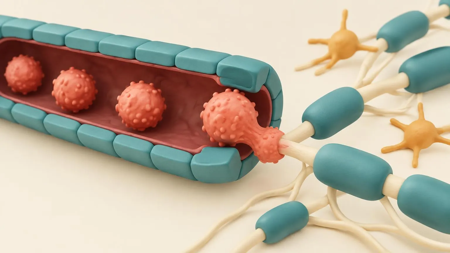
The brain and spinal cord are protected by the blood-brain barrier. The cells lining their small blood vessels are sealed to each other by tight junctions, wrapped in a basement membrane and surrounded by a second wall, the glia limitans, formed by the feet of astrocytes. An immune cell has to pass both walls to reach nerve tissue. Between them lies the perivascular space, a narrow sleeve around the vessel where immune cells can gather.
How an immune cell gets in
How an immune cell gets in
Step 1 of 6 · Activation in a lymph node.
What changes later in the disease
Early in MS, most new lesions come from waves of cells crossing from the blood, and medicines that stop this entry or remove the cells prevent most of them. As the disease goes on, part of the inflammation settles behind a barrier that has closed again. Microglia stay switched on at the edges of old lesions, and B cells gather in the membranes over the brain. Medicines that work in the blood reach this inflammation poorly, which is one reason progressive MS responds less to them. Lesson 3.2 and the frontier Why MS progresses follow this story.
Check yourself
What we know
- New lesions begin when immune cells cross from the blood into the brain or spinal cord.
- Blocking that entry, or removing B cells, prevents most new lesions in relapsing MS.
- Active lesions contain expanded families of CD8 T cells, and regulatory T cells work less well in MS.
What we don't know
- Which targets the T cells and B cells in MS recognise. No single target has been confirmed.
- Why the barrier opens at particular places and times.
- What keeps inflammation going behind a closed barrier, and how to reach it.
In the research now
Loading current research…
Key terms
- Macrophage
- A large immune cell that swallows debris, including stripped myelin. In the brain it comes from blood monocytes.
- Cytokine
- A signalling protein that immune cells release to direct other cells.
- HLA
- Human leucocyte antigen: molecules that display protein fragments to T cells. Different versions change MS risk.
- CD4 and CD8
- Markers that divide T cells into helpers (CD4), which direct responses, and cytotoxic cells (CD8), which kill.
- Regulatory T cell
- A T cell that holds other immune cells in check and helps prevent autoimmunity.
- Clonal expansion
- Many copies of one immune cell, made after it meets its target.
- Integrin
- A grip molecule on immune cells that lets them stick to vessel walls. Natalizumab blocks α4 integrin.
- Perivascular space
- The narrow sleeve around a blood vessel in the brain, between the vessel wall and the brain tissue.
- Glia limitans
- The inner wall of the blood-brain barrier, formed by the feet of astrocytes.
- PML
- Progressive multifocal leukoencephalopathy: a rare, serious brain infection by the JC virus, linked to some MS medicines.
- Blood-brain barrier
- The tight lining of the small blood vessels of the brain and spinal cord, which keeps most immune cells and large molecules out.
- T cell
- A white blood cell that recognises a specific target and directs or carries out an immune attack.
- B cell
- A white blood cell that can present targets to T cells, release inflammatory signals and mature into antibody-making cells. Some B-cell-targeting medicines are highly effective MS treatments.
- Plasma cell
- An antibody-making cell derived from a B cell. Most plasma cells do not carry CD20, so anti-CD20 medicines do not directly target them.
- Microglia
- The resident immune cells of the brain and spinal cord. In MS they sustain slow inflammation at the edges of chronic lesions.
- Astrocyte
- A star-shaped support cell of the brain and spinal cord. In AQP4-positive NMOSD, antibodies target a protein on astrocytes.
- Lesion (plaque)
- An area of damage where myelin has been lost and the tissue scarred.
- Relapse (attack)
- New or worsening symptoms lasting at least 24 hours, without fever or infection to explain them.
- Oligoclonal bands
- Distinct bands of antibodies found in spinal fluid but not matched in blood, showing antibody production inside the nervous system. They are common in MS but are not specific to it.
- Epstein-Barr virus (EBV)
- A common herpes virus that causes glandular fever. Infection appears to be almost necessary for MS, but most infected people never develop it.
- HLA-DRB1*15:01
- A version of an immune gene that is the strongest single genetic risk factor for MS.
- Anti-CD20
- Medicines that remove B cells by attaching to the CD20 protein on their surface, such as ocrelizumab and ofatumumab.
- S1P modulator
- A medicine that keeps immune cells in the lymph nodes by blocking the S1P signal they follow to leave, such as fingolimod.
- Phase 1, 2 and 3
- The stages of testing in people: safety and dose, then an early sign of effect, then a large test of benefit.
References
- Dendrou CA, Fugger L, Friese MA. Immunopathology of multiple sclerosis. Nat Rev Immunol 2015.https://doi.org/10.1038/nri3871
- Babbe H, et al. Clonal expansions of CD8+ T cells dominate the T cell infiltrate in active multiple sclerosis lesions as shown by micromanipulation and single cell polymerase chain reaction. J Exp Med 2000.https://doi.org/10.1084/jem.192.3.393
- Viglietta V, et al. Loss of functional suppression by CD4+CD25+ regulatory T cells in patients with multiple sclerosis. J Exp Med 2004.https://doi.org/10.1084/jem.20031579
- Hauser SL, et al. B-cell depletion with rituximab in relapsing-remitting multiple sclerosis. N Engl J Med 2008.https://doi.org/10.1056/NEJMoa0706383
- Engelhardt B, Ransohoff RM. Capture, crawl, cross: the T cell code to breach the blood-brain barriers. Trends Immunol 2012.https://doi.org/10.1016/j.it.2012.07.004
Next module · Lesions and pathology
How a lesion forms and evolves
A lesion is the basic unit of damage in MS. This lesson follows one from its first days to its possible endings: repair, a quiet scar, or a slowly burning edge that keeps growing.
Continue