How a lesion forms and evolves
What a lesion is
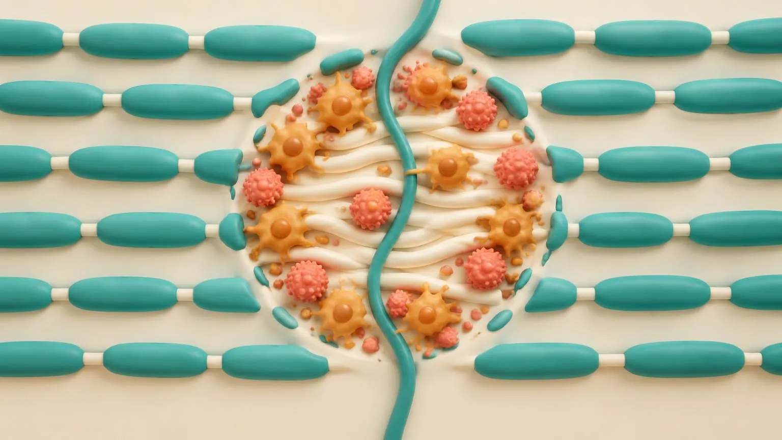
A lesion, or plaque, is an area of the brain, spinal cord or optic nerve where myelin has been destroyed. Lesions are what MRI scans show as bright spots and what gave the disease its name: the hardened patches, or sclerosis, that nineteenth-century doctors felt in the brains of people who had died with it.
Lesions are not scattered at random. They favour the tissue around the fluid-filled ventricles, the edge of the cortex, the brainstem, the spinal cord and the optic nerves, and most form around a small vein. That vein is the route by which immune cells arrive, and it can be seen on special MRI as the central vein sign (lesson 6.2).
From the first days to the endings
Lesion lifecycle
Day 1
Immune cells cross the vein wall and the barrier opens.
On MRI: Gadolinium leaks in and the lesion enhances.
A lesion begins when immune cells cross the wall of a small vein and the barrier around it opens. On MRI, the opened barrier lets the contrast agent gadolinium leak into the tissue, so the new lesion lights up, usually for a few weeks.
In the active phase, the whole lesion is filled with macrophages and microglia loaded with fragments of myelin they have stripped and swallowed. Oligodendrocytes die, and axons are cut. Damage to axons is greatest where inflammation is greatest: in one study active lesions held on average more than 11,000 cut axons per cubic millimetre.
Over the following weeks and months the inflammation settles. Precursor cells move in from the surrounding tissue, and the lesion heads towards one of three endings, often a mixture of them.
- Repaired. New oligodendrocytes wrap the bare axons in new, thinner myelin. Under the microscope the area looks paler than normal tissue and is called a shadow plaque.
- Inactive. The area stays without myelin. Few immune cells remain, astrocytes fill it with scar tissue, and many of its axons are gone.
- Smouldering. The centre is burnt out, but a rim of activated microglia and macrophages at the edge keeps stripping myelin very slowly, so the lesion creeps outwards for years. Lesson 3.2 is about these lesions.
How pathologists classify lesions
Pathologists sort lesions by their activity. The current classification separates active lesions, mixed active and inactive lesions (the smouldering ones), and inactive lesions, and notes whether myelin is still being stripped and whether the area has been remyelinated.
The largest study of lesion types examined 2,476 white matter plaques from 120 people with MS. Active plaques dominated early in the disease and in relapsing MS, and their number fell quickly with disease duration. Smouldering plaques were found frequently and almost only in progressive MS. At around 47 years of age, active and inactive plaques were in balance and smouldering plaques began to peak. Men had a higher proportion of smouldering plaques than women. The study suggests that MS moves over time from fresh lesions, which cause relapses, to slowly expanding old ones.
Remyelination: how often repair works
Repair happens in MS, but it varies greatly between people. In a study of 51 autopsies, one person in five had extensive repair, with 60 to 96 per cent of their total lesion area remyelinated, and these people included some with progressive MS. Others had very little.
Precursor cells are present in many lesions that never repair, so the problem is less often a lack of cells than their failure to mature into oligodendrocytes. Age is one reason. In rodents, older precursor cells respond less to signals telling them to mature, and fasting or metformin restored their ability to repair myelin. That finding led to trials in people, which the frontier Repair and protection follows.
Lesions and symptoms
A lesion causes symptoms only when it damages a pathway that matters and the brain cannot work around it. Many new lesions seen on MRI cause no symptoms at all, which is why scans find activity that people do not notice and why trials count new lesions as well as relapses. The opposite also happens. A small lesion in the spinal cord or brainstem can cause a large deficit.
Check yourself
What we know
- Lesions form around small veins when immune cells enter, and active lesions cut many axons.
- Lesions end repaired, inactive or smouldering, and the balance shifts towards smouldering lesions in progressive MS.
- Repair is extensive in some people and nearly absent in others, and ageing precursor cells are one reason.
What we don't know
- Why one lesion repairs fully while its neighbour becomes a scar.
- What decides whether a lesion keeps a smouldering edge.
- Whether medicines that promote repair can restore function in people. Early trials have shown repair can be measured, but none has yet shown a lasting gain in everyday function.
In the research now
Loading current research…
Key terms
- Gadolinium
- A contrast agent injected before an MRI scan. It leaks into lesions where the barrier is open.
- Shadow plaque
- A lesion that has been repaired with new, thinner myelin, so it looks paler than normal tissue.
- Macrophage
- A large immune cell that swallows debris, including stripped myelin. In the brain it comes from blood monocytes.
- Smouldering lesion
- A chronic lesion with a burnt-out centre and an active edge that slowly expands. Also called a chronic active or mixed active and inactive lesion.
- Myelin
- The fatty sheath wrapped around many nerve fibres in the brain and spinal cord. It lets signals jump quickly along the fibre. MS damages it.
- Axon (nerve fibre)
- The long extension of a nerve cell that carries its signal. Losing axons, rather than myelin alone, is thought to cause most lasting disability.
- Oligodendrocyte
- The cell in the brain and spinal cord that makes myelin. One oligodendrocyte can wrap many axons.
- Remyelination
- The brain's own repair of lost myelin, by new oligodendrocytes. It happens in MS but often falls short.
- Microglia
- The resident immune cells of the brain and spinal cord. In MS they sustain slow inflammation at the edges of chronic lesions.
- Astrocyte
- A star-shaped support cell of the brain and spinal cord. In AQP4-positive NMOSD, antibodies target a protein on astrocytes.
- Lesion (plaque)
- An area of damage where myelin has been lost and the tissue scarred.
- T2 lesion
- A lesion visible as a bright spot on a T2-weighted MRI sequence. New or enlarging T2 lesions, compared with an earlier scan, can show activity since that scan.
- Central vein sign
- A small vein running through the centre of a lesion on MRI. It can support an MS diagnosis in specified circumstances under the 2024 McDonald criteria.
- Relapse (attack)
- New or worsening symptoms lasting at least 24 hours, without fever or infection to explain them.
References
- Trapp BD, et al. Axonal transection in the lesions of multiple sclerosis. N Engl J Med 1998.https://doi.org/10.1056/NEJM199801293380502
- Kuhlmann T, et al. An updated histological classification system for multiple sclerosis lesions. Acta Neuropathol 2017.https://doi.org/10.1007/s00401-016-1653-y
- Frischer JM, et al. Clinical and pathological insights into the dynamic nature of the white matter multiple sclerosis plaque. Ann Neurol 2015.https://doi.org/10.1002/ana.24497
- Patrikios P, et al. Remyelination is extensive in a subset of multiple sclerosis patients. Brain 2006.https://doi.org/10.1093/brain/awl217
- Neumann B, et al. Metformin restores CNS remyelination capacity by rejuvenating aged stem cells. Cell Stem Cell 2019.https://doi.org/10.1016/j.stem.2019.08.015
Next · Lesson 2 of 2
Smouldering inflammation and the cortex
Inflammation in MS does not only arrive in bursts from the blood. Part of it settles inside the brain, at the edges of old lesions and in the membranes over the cortex, and keeps working slowly. It is thought to drive much of progression.
Continue