Smouldering inflammation and the cortex
Inflammation behind a closed barrier
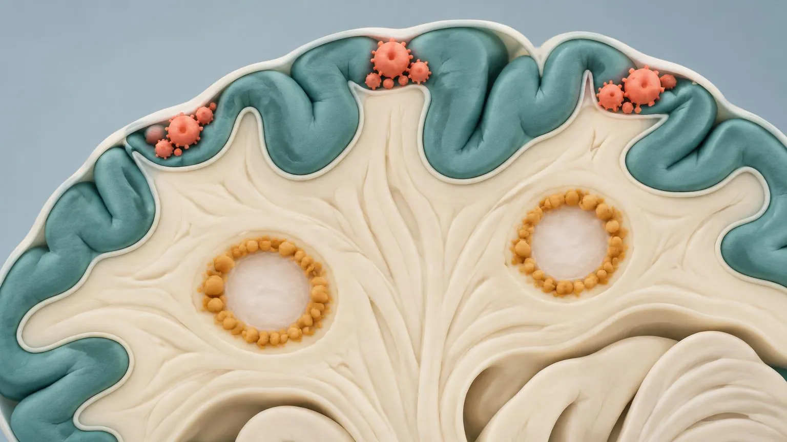
In relapsing MS, most damage comes from new lesions, each opened by a wave of cells from the blood. In progressive MS, the picture changes. Fewer lesions enhance on MRI, yet tissue keeps being lost. Studies of brain tissue showed why: inflammation persists inside the brain behind a barrier that has largely closed, at the edges of old lesions, across large areas of white matter that look normal on scans, and in the membranes over the cortex. This is called compartmentalised or smouldering inflammation.
Lesions with a burning edge
A smouldering lesion has a burnt-out centre and a rim of activated microglia and macrophages that keeps stripping myelin very slowly. Axons are lost at this edge too. In the 1998 study of cut axons, the edges of such lesions held more than 3,000 cut axons per cubic millimetre, against fewer than 900 in their centres.
The microglia at the rim contain iron, and iron disturbs the magnetic field in a way that susceptibility-weighted MRI can detect. These lesions therefore appear with a dark rim, and are called paramagnetic rim lesions. In a study of 209 people with MS at the US National Institutes of Health, 56 per cent had at least one rim lesion, including people on effective treatment. Those with four or more reached motor and cognitive disability at a younger age. In 23 people followed with yearly scans for ten years or more, lesions with rims stayed the same size or grew by 2.2 per cent a year, while lesions without rims shrank by 3.6 per cent a year.
A second method finds slowly expanding lesions on ordinary MRI, by measuring how much existing lesions grow between scans. In large trial datasets, people with primary progressive MS had more of them than people with relapsing MS. The two markers overlap only partly. In one group of 61 people, 92 per cent had at least one slowly expanding lesion and 56 per cent at least one rim lesion, and those with both kinds worsened faster on the EDSS than those with slowly expanding lesions alone.
Under the microscope
On MRI
The cortex and the membranes over it
MS damages grey matter as well as white matter, above all the cortex. Many cortical lesions lie just under the brain's surface and spread along it, beneath the meninges, the membranes that cover the brain. These lesions are hard to see on standard MRI, so they were underestimated for decades.
In the meninges, B cells, T cells and plasma cells can gather into structures that resemble the follicles of a lymph node. In brain tissue from 123 people with secondary progressive MS, such follicle-like structures were found in 40 per cent. Their presence went with more inflammation in the meninges, more activated microglia, more cortical demyelination and a more severe course of the disease. One explanation is that cells in the meninges release substances that diffuse into the cortex below and damage it from the surface inwards.
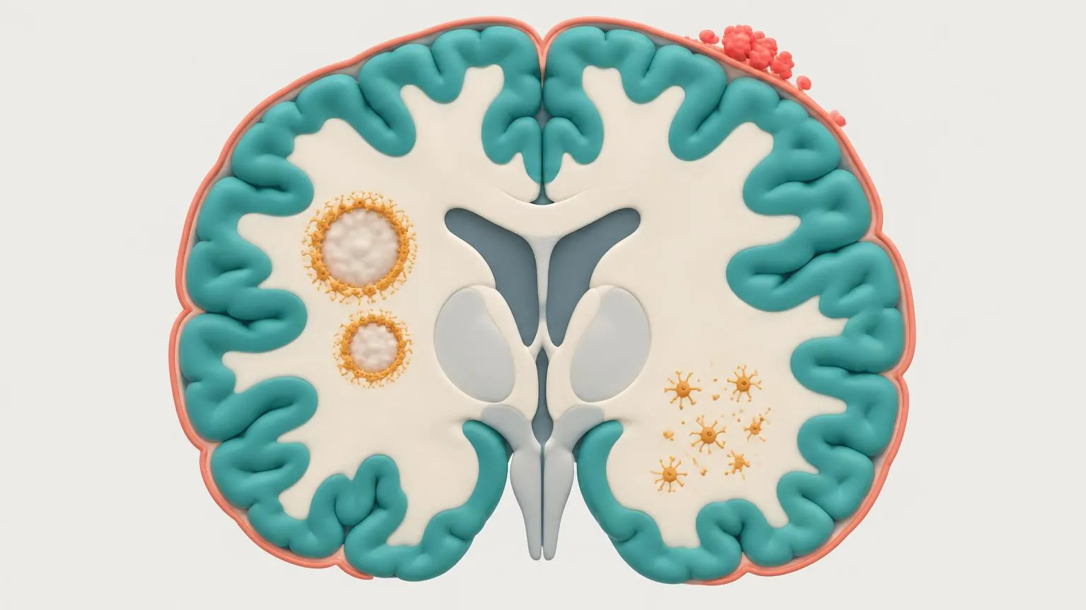
- Edges of old lesions
Activated microglia at the rim keep stripping myelin. Seen on MRI as rim lesions and slowly expanding lesions.
- The meninges
Immune cells gather in the membranes over the brain, sometimes in follicle-like clusters.
- The cortex
Lesions spread under the brain's surface, near inflamed meninges. Hard to see on standard MRI.
- Normal-looking white matter
Diffuse injury to axons and widespread microglial activation, invisible on standard scans.
Damage spread across the brain
Away from lesions, white matter that looks normal on MRI is not normal under the microscope. It shows diffuse injury to axons with widespread activation of microglia. A study of 52 brains found this diffuse injury and cortical demyelination mainly in primary and secondary progressive MS, and only a weak link between the number of focal white matter lesions and either kind of damage. This is part of the reason the number of lesions on a scan predicts disability poorly (lesson 6.2).
What this means for treatment
Most MS medicines act in the blood or lymph nodes and reach the brain poorly, so they do little against inflammation that has settled behind the barrier. Medicines that enter the brain and act on microglia and B cells there are the current test of the idea. In the HERCULES trial, 1,131 people with secondary progressive MS without relapses received tolebrutinib, a BTK inhibitor that enters the brain, or placebo. After a median of 133 weeks, 22.6 per cent of those on tolebrutinib had confirmed disability progression against 30.7 per cent on placebo, a hazard ratio of 0.69. Liver enzyme rises above three times normal occurred in 4.0 per cent of people on tolebrutinib and 1.6 per cent on placebo. The live box below shows the other trials of this idea and their results.
Check yourself
What we know
- Inflammation persists inside the brain in progressive MS, at the edges of old lesions, in the cortex and in the meninges.
- Rim lesions and slowly expanding lesions can be seen on MRI in living people, and both go with faster worsening.
- A brain-penetrant BTK inhibitor slowed disability in secondary progressive MS without relapses in one phase 3 trial.
What we don't know
- What keeps microglia active at a lesion's edge for years.
- How much of progression comes from smouldering inflammation and how much from ageing and failed repair.
- Whether shrinking rim lesions or slowing lesion growth on MRI will translate into less disability for individuals.
In the research now
Loading current research…
Key terms
- Macrophage
- A large immune cell that swallows debris, including stripped myelin. In the brain it comes from blood monocytes.
- Smouldering lesion
- A chronic lesion with a burnt-out centre and an active edge that slowly expands. Also called a chronic active or mixed active and inactive lesion.
- Slowly expanding lesion
- A lesion that grows slowly between MRI scans, a marker of smouldering inflammation.
- Meninges
- The membranes that cover the brain and spinal cord.
- Cortical lesion
- A lesion in the cortex, the outer grey matter of the brain. Often hard to see on standard MRI.
- Myelin
- The fatty sheath wrapped around many nerve fibres in the brain and spinal cord. It lets signals jump quickly along the fibre. MS damages it.
- Axon (nerve fibre)
- The long extension of a nerve cell that carries its signal. Losing axons, rather than myelin alone, is thought to cause most lasting disability.
- T cell
- A white blood cell that recognises a specific target and directs or carries out an immune attack.
- B cell
- A white blood cell that can present targets to T cells, release inflammatory signals and mature into antibody-making cells. Some B-cell-targeting medicines are highly effective MS treatments.
- Plasma cell
- An antibody-making cell derived from a B cell. Most plasma cells do not carry CD20, so anti-CD20 medicines do not directly target them.
- Microglia
- The resident immune cells of the brain and spinal cord. In MS they sustain slow inflammation at the edges of chronic lesions.
- Lesion (plaque)
- An area of damage where myelin has been lost and the tissue scarred.
- Central vein sign
- A small vein running through the centre of a lesion on MRI. It can support an MS diagnosis in specified circumstances under the 2024 McDonald criteria.
- Relapse (attack)
- New or worsening symptoms lasting at least 24 hours, without fever or infection to explain them.
- EDSS
- Expanded Disability Status Scale, from 0 (normal examination) to 10, the main disability measure in trials. Its middle range depends heavily on walking.
- Confirmed disability progression
- A rise in EDSS meeting a stated threshold and still present at a check, often three or six months later. The threshold and confirmation rules vary by trial.
- McDonald criteria
- The international criteria for diagnosing MS, last revised in 2024 and published in 2025.
- BTK inhibitor
- A medicine that blocks Bruton’s tyrosine kinase, an enzyme involved in signalling in B cells and microglia. How well it enters the brain varies between medicines.
- Placebo
- A dummy treatment that looks like the real one, used so that trial results reflect the medicine itself.
- Phase 1, 2 and 3
- The stages of testing in people: safety and dose, then an early sign of effect, then a large test of benefit.
- Hazard ratio
- A comparison of the event rate over time between groups. A hazard ratio of 0.76 means the estimated hazard was 24 per cent lower in one group during the study. It does not necessarily mean 24 per cent fewer people had the event.
References
- Dendrou CA, Fugger L, Friese MA. Immunopathology of multiple sclerosis. Nat Rev Immunol 2015.https://doi.org/10.1038/nri3871
- Trapp BD, et al. Axonal transection in the lesions of multiple sclerosis. N Engl J Med 1998.https://doi.org/10.1056/NEJM199801293380502
- Absinta M, et al. Association of chronic active multiple sclerosis lesions with disability in vivo. JAMA Neurol 2019.https://doi.org/10.1001/jamaneurol.2019.2399
- Elliott C, et al. Slowly expanding/evolving lesions as a magnetic resonance imaging marker of chronic active multiple sclerosis lesions. Mult Scler 2019.https://doi.org/10.1177/1352458518814117
- Calvi A, et al. Relationship between paramagnetic rim lesions and slowly expanding lesions in multiple sclerosis. Mult Scler 2023.https://doi.org/10.1177/13524585221141964
- Howell OW, et al. Meningeal inflammation is widespread and linked to cortical pathology in multiple sclerosis. Brain 2011.https://doi.org/10.1093/brain/awr182
- Kutzelnigg A, et al. Cortical demyelination and diffuse white matter injury in multiple sclerosis. Brain 2005.https://doi.org/10.1093/brain/awh641
- Fox RJ, et al. Tolebrutinib in nonrelapsing secondary progressive multiple sclerosis. N Engl J Med 2025.https://doi.org/10.1056/NEJMoa2415988
Next module · Attacks and progression
Types, courses and the spectrum
The main courses of MS, the rarer forms, and why experts now describe one disease along a spectrum.
Continue